How TMS Therapy Works: A Neurology Explainer for Patients and Families
Patients considering transcranial magnetic stimulation routinely ask two questions: *what does the device actually do to my brain?* and *why does that change my depression?* Both have clear, well-studied answers.
Article body
Patients considering transcranial magnetic stimulation routinely ask two questions: what does the device actually do to my brain? and why does that change my depression? Both have clear, well-studied answers. This article walks through the physics of the magnetic pulse, the neural circuit being targeted, and the cumulative biological effects that produce antidepressant response over a standard six-to-nine-week course. The intent is patient and family literacy — not a substitute for the clinical consultation, which determines candidacy and protocol.
The basic physics: a focused magnetic pulse, not electricity through the brain
Transcranial magnetic stimulation uses a copper coil held against the scalp to generate a brief, focused magnetic field — approximately 1.5 to 2 Tesla, comparable in strength to an MRI scanner — over roughly 100 to 300 microseconds.1 This rapidly changing magnetic field passes painlessly through the skull (bone is essentially transparent to magnetic fields) and induces a small electrical current in the cortical tissue directly beneath the coil. That induced current depolarizes neurons in a focal region of cortex, roughly 2 to 3 cm in diameter.
Two clarifications are useful:
- TMS is not ECT. Electroconvulsive therapy delivers electrical current through scalp electrodes under anesthesia, intentionally inducing a generalized seizure. TMS induces a small, focal current via a magnetic field, in awake, alert patients — and is specifically designed not to provoke seizure. (Seizure is a rare adverse event, estimated at fewer than 1 in 60,000 sessions when published safety guidelines are followed.)2
- TMS does not deliver any drug or electrical current systemically. Nothing is injected, infused, or swallowed. The effect is confined to the cortical region under the coil and the networks downstream of it.
The first device cleared in the United States for this indication — and still the most widely deployed in outpatient psychiatry — is the NeuroStar Advanced Therapy System, FDA-cleared under 510(k) K061053 in 2008 for major depressive disorder in adults who have failed to achieve satisfactory improvement from prior antidepressant medication.3 Other FDA-cleared systems (e.g., BrainsWay Deep TMS, MagVenture) use different coil geometries but share the underlying mechanism described below.
The target: the left dorsolateral prefrontal cortex
For depression, the treatment coil is positioned over the left dorsolateral prefrontal cortex (DLPFC) — a region on the upper-front-left of the head, roughly 5 to 6 cm anterior to the motor cortex. The DLPFC is selected for two reasons grounded in clinical neuroscience.
First, functional imaging consistently shows hypoactivity in the left DLPFC in patients with major depressive disorder. The DLPFC participates in executive function, attention regulation, emotional appraisal, and cognitive control of negative affect — all functions clinically impaired in depression.
Second, and more importantly, the DLPFC is anti-correlated in network terms with the subgenual anterior cingulate cortex (sgACC) — a deeper limbic region that shows abnormally high activity in depression and that is a known node of the brain's "depression network." Fox and colleagues (2012) demonstrated that the more strongly a DLPFC stimulation site is anti-correlated with the sgACC, the better the antidepressant response.4 In other words: TMS targets a superficial cortical site, but its therapeutic effect propagates through known network connections to deeper regions that surface stimulation cannot reach directly.
The motor threshold — the lowest stimulator output that produces a visible muscle twitch when the coil is over the motor cortex — is measured at the first treatment and used to set treatment intensity, typically at 120% of motor threshold.
What changes in the brain over a course of treatment
A single TMS pulse depolarizes neurons under the coil. A repetitive pulse train — repetitive TMS (rTMS) — produces effects that outlast the stimulation itself, on the order of minutes to hours after a single session and weeks to months after a complete course. The leading mechanistic explanation is that rTMS induces synaptic plasticity analogous to long-term potentiation (LTP) and long-term depression (LTD), the cellular mechanisms underlying learning and memory.1
High-frequency stimulation (10 Hz over the left DLPFC, the standard depression protocol) is excitatory — it strengthens synaptic connections and increases cortical excitability in the targeted region. Over a full course, measurable changes include:
- Increased glucose metabolism and cerebral blood flow in the left DLPFC
- Normalization of functional connectivity between DLPFC and sgACC
- Changes in BDNF (brain-derived neurotrophic factor), suggesting downstream neuroplastic effects
- Modulation of monoaminergic systems (serotonin, dopamine, norepinephrine) — the same systems that oral antidepressants influence, but reached via cortical circuitry rather than via the bloodstream
This is why a standard course is approximately 36 sessions over six to nine weeks. The cumulative neuroplastic effect — not any single session — produces clinical response.
The two main protocols: 10 Hz rTMS and intermittent theta burst (iTBS)
Two FDA-cleared protocols are in routine clinical use:
- 10 Hz rTMS — the original NeuroStar protocol. Pulses delivered at 10 Hz in 4-second trains with 26-second inter-train intervals, for approximately 3,000 pulses per session over roughly 37 minutes.
- Intermittent theta burst stimulation (iTBS) — a patterned protocol delivering 600 pulses in approximately 3 minutes by mimicking endogenous theta-rhythm bursts. The THREE-D non-inferiority trial demonstrated that iTBS produces equivalent response and remission rates to standard 10 Hz rTMS in TRD, with dramatically shorter session times.5
The choice between protocols is made by the treating psychiatrist based on tolerability, scheduling, and prior response history. Either is appropriate for most patients with TRD; both qualify for insurance coverage under the same indication.
What patients feel during a session
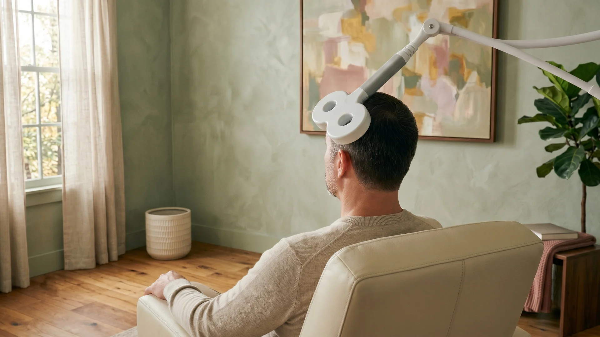
The most common physical sensations during a TMS session are a tapping or clicking sensation on the scalp under the coil, mild contraction of the scalp and facial muscles near the stimulation site, and the audible clicking of the device itself (ear protection is provided). Patients are awake, seated in a treatment chair, and can read, listen to audio, or talk during treatment. No sedation is used. Most patients drive themselves to and from sessions and return to work or normal activity immediately afterward.
Common, transient side effects include scalp discomfort at the treatment site and headache, both typically mild; tolerability generally improves over the first one to two weeks of treatment as the scalp accommodates.67 Serious adverse events — most notably seizure — are rare when established safety guidelines are followed.8
Key takeaways
- TMS uses a focused magnetic field to induce a small electrical current in a focal region of cortex; nothing is injected and no electrical current passes through the skull directly.
- The target for depression is the left dorsolateral prefrontal cortex, selected because its network anti-correlation with the subgenual cingulate predicts antidepressant response.
- A standard course is approximately 36 sessions over six to nine weeks; therapeutic effect comes from cumulative synaptic plasticity, not any single pulse.
- Two FDA-cleared protocols — 10 Hz rTMS and iTBS — produce equivalent outcomes; iTBS sessions are shorter (≈3 minutes vs. ≈37 minutes).
- Patients remain awake and unsedated; the most common side effects are scalp discomfort and headache, typically transient.
For patients in Anaheim, Orange County, and the broader 30-mile radius who want to understand whether TMS therapy is appropriate for their treatment history, our psychiatrists conduct the candidacy evaluation in a single consultation. Most patients begin by verifying insurance coverage so the full picture — clinical fit and financial fit — is clear from the outset.
Sources / Further reading
Klomjai W, Katz R, Lackmy-Vallée A. Basic principles of transcranial magnetic stimulation (TMS) and repetitive TMS (rTMS). Ann Phys Rehabil Med. 2015;58(4):208–213. ↩ ↩
Lerner AJ, Wassermann EM, Tamir DI. Seizures from transcranial magnetic stimulation 2012–2016: results of a survey of active laboratories and clinics. Clin Neurophysiol. 2019;130(8):1409–1416. ↩
U.S. Food and Drug Administration. 510(k) Premarket Notification K061053, NeuroStar TMS Therapy System (Neuronetics, Inc.), cleared 2008 for treatment of major depressive disorder in adult patients who have failed to achieve satisfactory improvement from prior antidepressant medication. ↩
Fox MD, Buckner RL, White MP, Greicius MD, Pascual-Leone A. Efficacy of transcranial magnetic stimulation targets for depression is related to intrinsic functional connectivity with the subgenual cingulate. Biol Psychiatry. 2012;72(7):595–603. ↩
Blumberger DM, Vila-Rodriguez F, Thorpe KE, et al. Effectiveness of theta burst versus high-frequency repetitive transcranial magnetic stimulation in patients with depression (THREE-D): a randomised non-inferiority trial. Lancet. 2018;391(10131):1683–1692. ↩
Carpenter LL, Janicak PG, Aaronson ST, et al. Transcranial magnetic stimulation (TMS) for major depression: a multisite, naturalistic, observational study of acute treatment outcomes in clinical practice. Depress Anxiety. 2012;29(7):587–596. ↩
Janicak PG, O'Reardon JP, Sampson SM, et al. Transcranial magnetic stimulation in the treatment of major depressive disorder: a comprehensive summary of safety experience from acute exposure, extended exposure, and during reintroduction treatment. J Clin Psychiatry. 2008;69(2):222–232. ↩
Rossi S, Antal A, Bestmann S, et al. Safety and recommendations for TMS use in healthy subjects and patient populations, with updates on training, ethical and regulatory issues: Expert Guidelines. Clin Neurophysiol. 2021;132(1):269–306. ↩